
Fig. 1
- ID
- ZDB-FIG-250417-22
- Publication
- Xu et al., 2024 - Single-cell RNA sequencing reveals the heterogeneity and interactions of immune cells and Müller glia during zebrafish retina regeneration
- Other Figures
- All Figure Page
- Back to All Figure Page
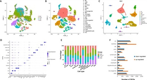
Characterization of zebrafish retinal cell types by scRNAseq.(A–C) Clustering of retinal cells in intact and stab-injured zebrafish retinas by t-SNE and UMAP. Cluster 3 expressing both AC and RGC markers was designated as AC/RGC or AC_RGC. (D) Cell type–specific marker genes. (E) Cell type composition of retinal cells from all four time points tested. (F) The number of DEGs in each cell type in the uninjured and injured retina. Uninjured group: 0 d; injured group: 12 hours, 1 day, and 2 days samples combined. DEGs were defined using the following criteria: P < 0.01, log2FC ≥ 0.26. AC: Amacrine cells; BC: bipolar cell; DEGs; differentially expressed genes; HC: horizontal cells; MG: Müller glia; RGC: retinal ganglion cells; RPE: retinal pigment epithelium; t-SNE: t-distributed stochastic neighbor embedding; UMAP: Uniform Manifold Approximation and Projection. |