FIGURE

Fig 8

ID
ZDB-FIG-250321-16
Publication
Nonarath et al., 2025 - The USH3A causative gene clarin1 functions in Müller glia to maintain retinal photoreceptors
Other Figures
All Figure Page
Back to All Figure Page
Fig 8

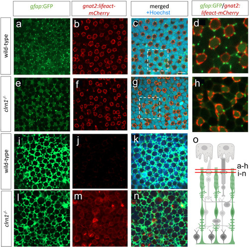
Interactions between Müller glia and cone photoreceptor cells are disorganized in clrn1-/-zebrafish.

En face images from Tg(gfap:GFP); Tg(gnat2:Lifeact-mCherry) 7 dpf transgenic zebrafish at the level of UV cone outer segments in either (a-d) wild-type or (e-h) clrn1-/- retina. Comparison of higher magnification images of (d) wild-type versus (h) clrn1-/- highlight abnormal interactions between Müller glia apical processes and cone calyceal processes within mutants. (i-n) En face images from Tg(gfap:GFP); Tg(gnat2:Lifeact-mCherry) 7 dpf transgenic zebrafish at the level of the OLM just apical of the adherens junctions, Müller glia processes encircle photoreceptors. Comparison of (i) wild-type versus (l) clrn1-/- highlight disorganization in mutants. At this image plane, actin is not well detected in (j) wild-types, but accumulates in (m) clrn1-/- cone photoreceptors. (o) Cartoon showing the imaged planes. Scale bar (c) = 5 µm. Schematic in (o) created in BioRender under License: https://BioRender.com/x12n534.

Expression Data

Expression Detail
Antibody Labeling
Phenotype Data

Phenotype Detail
Acknowledgments
This image is the copyrighted work of the attributed author or publisher, and ZFIN has permission only to display this image to its users. Additional permissions should be obtained from the applicable author or publisher of the image. Full text @ PLoS Genet.