|
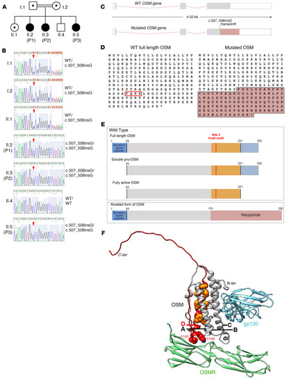
Identification of a homozygous OSM mutation in the patients. (A) Family pedigree. (B) Direct Sanger sequencing of the OSM gene in family members. Red arrowheads indicate the position of the 1 bp insertion causing a frameshift (c.507_508insG; Arg170AlafsTer124). (C) Schematic of the OSM gene showing the position of the frameshift leading to the production of a neo coding sequence (purple). (D) Protein sequences of the WT and mutated forms of OSM. Sequence in purple corresponds to the neopeptide generated by the frameshift. The red box highlights the FxxK motif critical to interact with OSMR and LIFR. (E) Domain architecture of the immature and mature forms of human OSM protein and the OSM mutated protein. Orange portion highlights the part that is missing in the mutated form of OSM and replaced by the neopeptide generated by the frameshift (in purple). Site 3 FxxK corresponds to the motif that interacts with OSMR and LIFR. (F) AlphaFold2 model of the 3D structure of WT human type II OSMR complex (OSM (UniProt P13725, aa 25-252) in complex with OSMR (UniProt Q99650, aa 141–330) and gp130 (UniProt P40189, aa 124–323). Details of the AF2 modeling are given in Supplemental Figure 6. The helices of the OSM 4-helix bundle are labeled. The phenylalanine and lysine of the FxxK motif are shown in a sphere representation (red) as well as hydrophobic amino acids of helix D (orange). The orange and red parts correspond to the missing domains in the OSMfs mutant created by the c.507_508insG variant.
|