FIGURE

Figure 1

ID
ZDB-FIG-250318-38
Publication
Garrigue et al., 2025 - Human Oncostatin M deficiency underlies an inherited severe bone marrow failure syndrome
Other Figures
All Figure Page
Back to All Figure Page
Figure 1

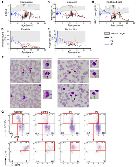
Evolution of hematologic values in the 3 patients, cytological analysis of bone marrow smears, and in vitro erythrocyte differentiation.

Variation of hemoglobin (g/dL) (A), hematocrit (%) (B), RBC (C), platelets (D), and neutrophil counts (E), in P1, P2, and P3 according to age. Gray zone indicates normal ranges in healthy individuals. The RBC counts were low in patients, even after RBC transfusion. (F) Cytological findings of bone marrow smears in P1 and P2. Irregular budding nucleus (red arrow), internuclear bridges (black arrow), acidophilic erythroblasts (green arrow), and polychromatophilic erythroblasts (orange arrow) were detected. Dysgranulopoiesis was observed in P1 as several metamyelocytes did not contain any granules (blue arrow). The slides were stained with Wright-Giemsa and H&E. Original magnification, ×100; ×175 (zoomed images). Abnormal cells are zoomed-in on the right. (G) In vitro differentiation of HSPCs into erythroblasts. Top: CD235a/CD45 staining after 7 days of culture in erythroid conditions of HSPCs from 2 healthy controls and patients P1 and P2. The red rectangle indicates the gating strategy on CD235a+CD45 erythroid cells. Bottom; CD36/CD71 staining of CD235a+CD45 cells. The red rectangle indicates CD36+CD71+ erythroblast cells.

Expression Data

Expression Detail
Antibody Labeling
Phenotype Data

Phenotype Detail
Acknowledgments
This image is the copyrighted work of the attributed author or publisher, and ZFIN has permission only to display this image to its users. Additional permissions should be obtained from the applicable author or publisher of the image. Full text @ Journal of Clin. Invest.