|
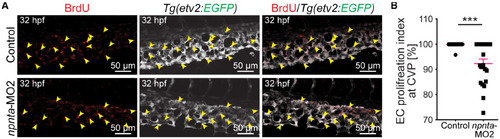
Decreased endothelial cell proliferation in the caudal vein plexus of npnta morphants. A Maximum projections of confocal images of 32 hpf whole‐mount embryos stained for 5‐bromo‐2′‐deoxyuridine (red; marking proliferating cells) and EGFP (pseudo color; labeling ECs). Arrowheads indicate 5‐bromo‐2′‐deoxyuridine+/EGFP+ cells. B, Quantitative analysis of EC proliferation at the caudal vein plexus. Graph representing the ratio of EGFP+/ethynyl‐2′‐deoxyuridine+ ECs relative to the total number of EGFP+ ECs at the CVP. A total of 24 control and 21 npnta morphants from 2 independent experiments were analyzed. The mean EC proliferative index for control embryos was considered as 100%. Statistical significance was determined by a 2‐tailed Student's t test. Data are mean±SEM. ***P≤0.001. CVP indicates caudal vein plexus; EC, endothelial cell; EGFP, enhanced green fluorescent protein; hpf, hours post fertilization; and MO2 spliced blocking morpholino.
|