
Fig. 4
- ID
- ZDB-FIG-250114-18
- Publication
- Horn et al., 2025 - Isotonic medium treatment limits burn wound microbial colonisation and improves tissue repair
- Other Figures
- All Figure Page
- Back to All Figure Page
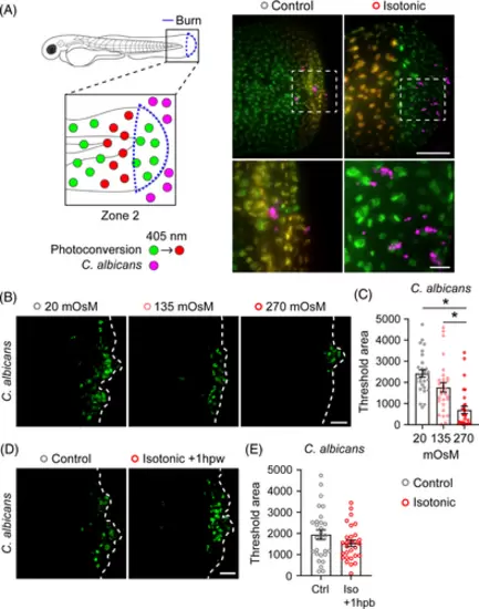
Keratinocyte dynamics enable microbial colonisation of burn wounded tissue. (A) Schematic of photoconversion experiment. Tg(Krt4-H2B-Dendra2) larvae were photoconverted to track zone 2 keratinocytes following exposure to far red-expressing C. albicans. Images show spatial localisation of C. albicans (magenta) relative to zone 2 keratinocytes (yellow) with the indicated treatment 1 hpb. Dashed box indicates region of inset shown below. Scale bar = 100 and 20 μm (inset). (B) C. albicans colonisation of wound area 24 hpb. Larvae were treated with medium supplemented with NaCl to a final concentration of 10 mOsm (Control), 135 mOsm or 270 mOsm (Isotonic). Dashed line indicates tailfin boundary. (C) Quantification of C. albicans colonisation from B. N = 29 larvae for each condition. (D) C. albicans colonisation of wound area 24 hpb. All larvae were burned in control medium, and treatment with isotonic medium began 1 hpb. Dashed line indicates tailfin boundary. (E) Quantification of C. albicans colonisation from D. N = 28 control and 29 isotonic treated larvae. Scale bars = 50 μm unless otherwise indicated. Asterisk indicates p < 0.05 by Kruskal–Wallis test (C) and independent t-test (E). |