|
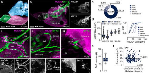
Neuroanatomy of the adult zebrafish intracardiac nervous system. a Color-coded presentation of the major regions of the adult zebrafish prototypic heart. b A representative whole-mount photomicrograph shows the general innervation pattern of the adult zebrafish heart with Zn-12 immunostaining (neuronal processes) and HuC/D (neuronal somata). The regions around the valves connecting the heart chambers hold the majority of the observed HuC/D+ neuronal somata (inserts i-iii). c Quantification of the proportion of HuC/D+ neurons found in different heart regions. d Regionalized quantification of the HuC/D+ cell soma size in the adult zebrafish heart. e Quantification of the total number of the HuC/D labeled neurons in the SAP area. f Analysis of the neuronal soma size in relation to relative distance from the sinoatrial valve. AVP atrioventricular plexus, BA bulbus arteriosus, CDF cumulative distribution frequency, HuC/D elav3&4, SAP sinoatrial plexus, SN sinus venosus, VBAP ventriculo-bulbus arteriosus plexus, Zn-12 neuronal cell surface marker (HNK-1). Data are presented as box plots showing the median with 25/75 percentile (box and line) and minimum-maximum (whiskers) and as mean ± SEM. For detailed statistics, see Supplementary Table 1. Source data are provided as a Source Data file.
|

