
Fig. 8
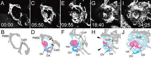
Formation of the CVP. Selected time-lapse images of a living Tg(flk1:EGFP)k7 embryo from 1.25 dpf (S6 Movie) (A, C, E, G, and I) and their schematic diagrams (B, D, F, H, and J). The time (hours:minutes) from the first frame is labeled in each image (A, C, E, G, and I). Rostral is facing right and dorsal is facing upward. To visualize CVP formation, only the selected slices from S5 Movie were projected. Ocular vessels in the schematic diagrams are colored (OA: pink, OV: sky blue, and CVP: light blue). Arrows in D indicate the rostral sprouts forming the CVP. Black arrowheads in H indicate the caudal sprouts forming the CVP. White arrowheads in F and H indicated the vascular plexus of the OV surrounding the OA. Asterisks in D, F and H indicates the new connection from the dorsal branch of the PMBC to the aCAC. |