
FIGURE
Fig. 1
Fig. 1
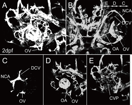
The primary ocular vasculature in zebrafish. (A–E) Ocular vascular anatomy of Tg(flk1:EGFP)k7 embryos at 2 dpf. Lateral (A, C, D, and E) and ventral (B) views. Lateral image of A divided into three parts: the superficial ocular (C) and hyaloid (D) vasculatures and the CVP (E). Panels C, D, and E in B indicate the projected region of each image. Arrows in B and D indicate the vascular plexus of the HA. Arrowheads in C indicate the IOC forming, which will connect the superficial ocular vasculature with the hyaloid vasculature. |
Expression Data
Expression Detail
Antibody Labeling
Phenotype Data
Phenotype Detail
Acknowledgments
This image is the copyrighted work of the attributed author or publisher, and
ZFIN has permission only to display this image to its users.
Additional permissions should be obtained from the applicable author or publisher of the image.
Full text @ PLoS One