
FIGURE
Fig. 2
Fig. 2
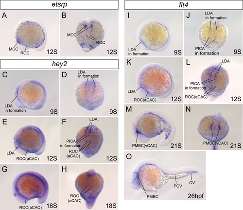
Arterial and venous cerebral angioblast clusters in zebrafish. Whole-mount in situ hybridization of WT embryos at 9S (C, D, I, and J), 12S (A, B, E, F, K, and L), 18S (G and H), 21S (M and N), and 26hpf (O). The expression of etsrp/etv2 (A and B), hey2 (C–H) and flt4 (I–O) genes was analyzed in both lateral view (A, C, E, G, I, K, M and O) and dorsal view of the cranial region (B, D, F, H, J, L and N). The differentiation of the aCAC and vCAC from the ROC and MOC was demonstrated. PCV: posterior cardinal vein. CV: caudal vein. |
Expression Data
Expression Detail
Antibody Labeling
Phenotype Data
Phenotype Detail
Acknowledgments
This image is the copyrighted work of the attributed author or publisher, and
ZFIN has permission only to display this image to its users.
Additional permissions should be obtained from the applicable author or publisher of the image.
Full text @ PLoS One