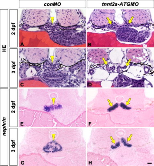
A tnnt2a-ATG morphant displays glomerulus disorganization similar to that found in the mpp5am520 mutants. Pronephric glomerular structure and nephrin mRNA expression are shown by hematoxylin-eosin stained section (A-F) and in situ hybridization with eosin-stained (G-J), respectively. In wild-type (A, C, E, G, I), a pair of glomerular primordia has already merged to form a single glomerulus (arrowheads) beneath the notochord at 2 dpf (A, G), 3 dpf (C, I), and 4 dpf (E). In tnnt2a-ATG morphants (B, D, F, H, J), a pair of glomerular primordia has retained their epithelial vesicular structure and remains unmerged at the midline in 2 dpf (arrows in B, H). Glomerular primordia have still not merged, and an extremely dilated dorsal aorta is interposed between glomerular primordia at 3 dpf (D) and 4 dpf (F). Bowman′s space is apparent in most cases of tnnt2a-ATG morphants (D, F). nephrin-expressing regions are moustache-like in shape and expanded in the direction of the pronephric tubule (arrows in H, I), as is the case in the mpp5a mutants. Scale bar = 10 μm.
|
