
- Title
-
gdf11 is required for pronephros/cloaca development through targeting TGF-β signaling
- Authors
- Tian, X., Yao, W., Tan, J., Hu, Z., Liu, J.
- Source
- Full text @ Sci. Rep.
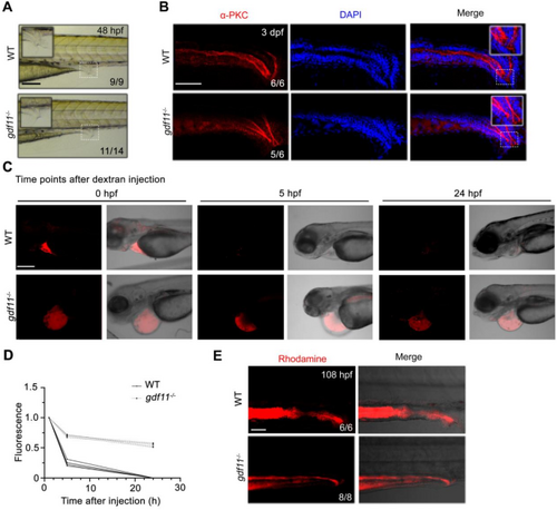
gdf11 knockout disrupts the function of the pronephros and cloaca in zebrafish. (A) Lateral view of pronephric tubules and cloaca at 36 hpf in WT and gdf11−/− mutants. Scale bar: 200 μm. (B) Lateral view of immunofluorescence images of α-PKC staining in both wild-type and gdf11 mutant embryos. Scale bar: 100 μm. (C,D) gdf11 mutant embryos exhibit slower clearance of dextran compared to WT embryos. Confocal images of embryos injected with 40 kDa rhodamine-dextran in the pericardial area immediately, and at 5–24 h post-injection (C). Quantification of fluorescence intensity (D). Scale bar: 50 μm. (E) Wild-type embryos rapidly secrete rhodamine dextran from the cloaca within minutes, while gdf11 mutants excrete only minimal dye. Scale bar: 50 μm. |
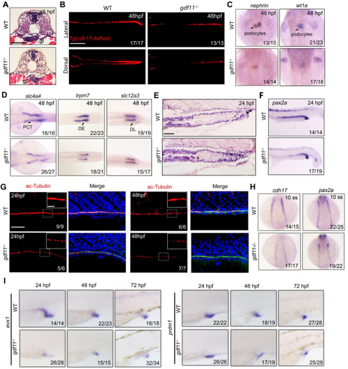
gdf11 is essential for pronephric tubule and cloaca organogenesis. (A) Hematoxylin-Eosin stained sections of WT and gdf11 mutant larvae at the level of the glomerulus and proximal tubules at 48 hpf. Scale bar: 50 μm. (B) The expression of cdh17 in WT and gdf11 mutants under Tg (cdh17-dsRed) background. Scale bar: 200 μm. (C) In situ hybridization of nephrin (left) and wt1a (right) in wild-type and gdf11 mutant embryos at 48 hpf. (D) WISH analysis in WT and gdf11 mutants at 48 hpf for slc4a4, trpm7, and slc12a3 probes. (E) H&E staining of the pronephric duct and cloaca region in WT and gdf11 mutants at 48 hpf. Scale bar: 200 μm. (F) In situ hybridization with pax2a in WT and gdf11 mutants at 24 hpf. (G) Confocal images showing cilia specification in WT and mutant embryos at 24 hpf and 48 hpf. Scale bar: 50 μm. (H) Comparative in situ hybridization of cdh17 and pax2a expression in WT and gdf11 mutants at 10 ss. (I) WISH analysis for evx1 and prdm1 in WT and gdf11 mutants from 24 hpf to 72 hpf. |
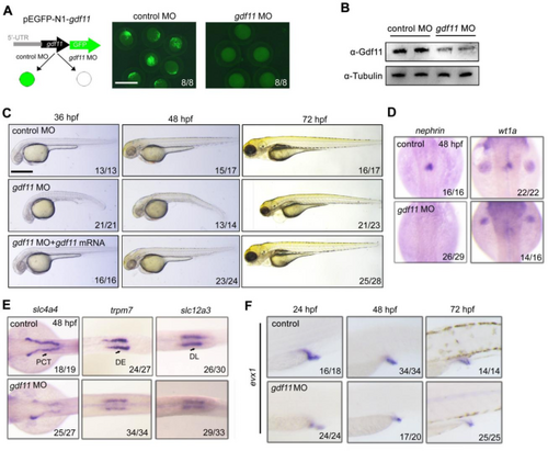
gdf11 knockdown disrupts pronephros and cloaca organogenesis. (A) Schematic representation of the plasmid used to test gdf11 MO efficiency. Scale bar: 500 μm. (B) Western blot showing a significant reduction of Gdf11 protein levels in gdf11 morphants. (C) Live imaging of control and gdf11 morphants, with or without gdf11 mRNA injection, from 36 hpf to 72 hpf. Scale bar: 300 μm. (D) Control and gdf11 morphant embryos harvested at 48 hpf were subjected to WISH for nephrin and wt1a expression. (E) ISH analysis using segment-specific probes for the pronephric tubules in control and gdf11 morphants at 48 hpf. (F) Lateral views of evx1 expression in control and gdf11 morphants from 24 hpf to 72 hpf. |
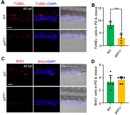
The apoptosis of pronephric tubules and cloaca was significantly decreased upon gdf11 deletion. (A,B) TUNEL assay showing decreased apoptosis in cells of the developing pronephric duct and cloaca in gdf11 mutants. The position marked by the dotted line in the figure is the pronephros and the cloaca. Scale bar: 50 μm. (C,D) Cell proliferation, indicated by BrdU staining, remains unchanged in both WT and gdf11 mutants. The position marked by the dotted line in the figure is the pronephros and the cloaca. Scale bar: 50 μm. |
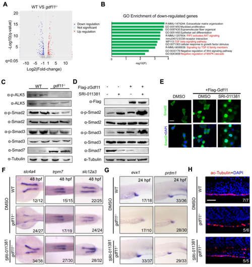
TGF-β signaling acts downstream of Gdf11 promoting pronephros and cloaca formation. (A) Volcano plot highlighting differentially expressed genes in WT and gdf11−/− pronephros. (B) Gene Ontology analysis of biological processes using down-regulated differentially expressed genes. (C) Western blot showing key proteins of the TGF-β/Smad signaling pathway in WT and gdf11−/− mutant pronephros/cloaca lysates. (D) Comparative expression of key TGF-β/Smad pathway proteins following Gdf11 overexpression or SRI-011381 treatment in HEK 293T cells. (E) Subcellular localization of Smad2 in Flag-Gdf11-infected cells with or without SRI-011381 treatment. Scale bar: 50 μm. (F) Partial rescue of pronephros developmental defects in gdf11 mutants by SRI-011381 treatment. (G) Partial rescue of cloaca developmental defects in gdf11 mutants by SRI-011381 treatment. (H) Partial rescue of cilia in pronephric duct in gdf11 mutants by SRI-011381 treatment. Scale bar: 50 μm. |
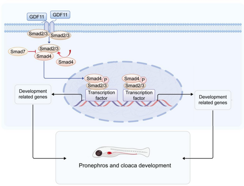
Schematic of TGF-β signaling downstream of Gdf11 in pronephros and cloaca organogenesis. Gdf11 enhances TGF-β signaling, supporting pronephros and cloaca development. This schematic highlights Gdf11’s essential role in zebrafish, illustrating the molecular mechanisms regulated by Gdf11. |