
- Title
-
The role of mip in the development of lens in zebrafish
- Authors
- He, M., Zhou, G., Lin, Q., Zhou, N.
- Source
- Full text @ Gene Expr. Patterns
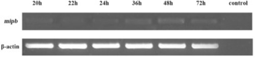
Mipb expression in zebrafish via RT-PCR. Mipb expression increased from 20 hpf to 72 hpf. EXPRESSION / LABELING:
|
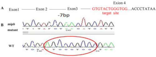
Mipb mutants resulted in the synthesis of a truncated protein (A) The fourth exon of mipb was selected as the target site for CRISPR/Cas9. (B) A 7-bp deletion was identified in exon 4 of mipb−/− by sequencing. |
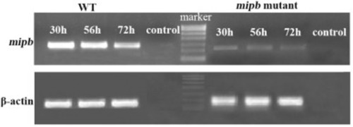
Analysis of mipb transcription in embryos via RT-PCR. The mRNA expression of mipb in mutants declined compared with that in WT fishes. EXPRESSION / LABELING:
PHENOTYPE:
|
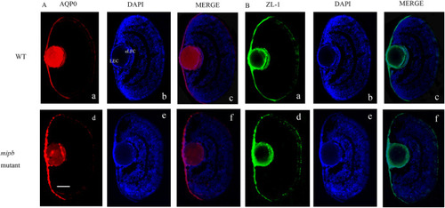
Distribution of MIP and ZL-1 protein in mutant lens (A) While mutant lens demonstrated a great decline in Mip protein (AQP0) expression compared to that in WT lens. (B) Diffuse distribution of ZL-1 positive signal was observed in the mutant lens while it was mainly located in the lens epithelial cells and elongating fiber cells in WT fish lens. AQP0: red, ZL-1: green; DAPI: blue. Scale bar = 25 μm. EXPRESSION / LABELING:
PHENOTYPE:
|
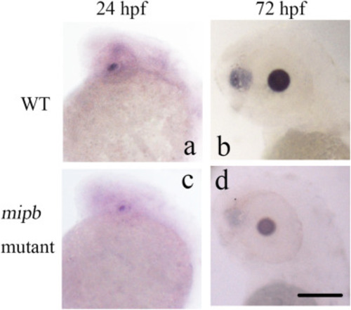
Bfsp2 expression analysis. Decreased expression of Bfsp2 in the mipb−/− fish lenses were found in situ hybridization. Scale bar = 50 μm. EXPRESSION / LABELING:
PHENOTYPE:
|
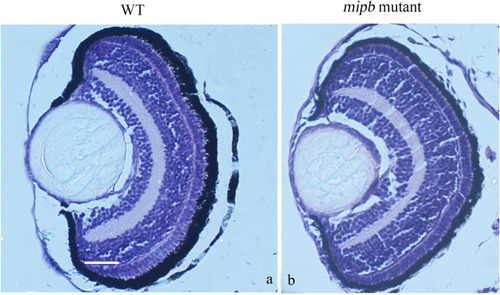
Histology of the mipb mutant eyes. The WT lens organization appeared as a sphere mass of lens fiber cells in the center and surrounded by a single layer of epithelial cell. The mipb−/− presented difference especially in the layer of epithelial cell from the WT eye morphology at 5 dpf. Scale bar = 25 μm. PHENOTYPE:
|
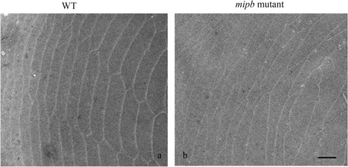
Lens fiber observed by TEM. Mutant lens fibers were disorganized and less uniform in shape. Scale bar = 2 μm. PHENOTYPE:
|
Reprinted from Gene expression patterns : GEP, 49, He, M., Zhou, G., Lin, Q., Zhou, N., The role of mip in the development of lens in zebrafish, 119330, Copyright (2023) with permission from Elsevier. Full text @ Gene Expr. Patterns