
FIGURE
Fig. 6
- ID
- ZDB-FIG-231228-29
- Publication
- He et al., 2023 - The role of mip in the development of lens in zebrafish
- Other Figures
- All Figure Page
- Back to All Figure Page
Fig. 6
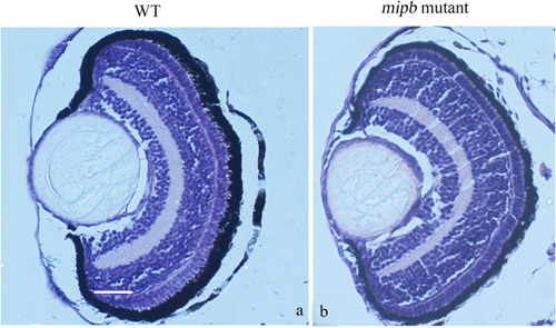
Histology of the mipb mutant eyes. The WT lens organization appeared as a sphere mass of lens fiber cells in the center and surrounded by a single layer of epithelial cell. The mipb−/− presented difference especially in the layer of epithelial cell from the WT eye morphology at 5 dpf. Scale bar = 25 μm. |
Expression Data
Expression Detail
Antibody Labeling
Phenotype Data
| Fish: | |
|---|---|
| Observed In: | |
| Stage: | Day 5 |
Phenotype Detail
Acknowledgments
This image is the copyrighted work of the attributed author or publisher, and
ZFIN has permission only to display this image to its users.
Additional permissions should be obtained from the applicable author or publisher of the image.
Reprinted from Gene expression patterns : GEP, 49, He, M., Zhou, G., Lin, Q., Zhou, N., The role of mip in the development of lens in zebrafish, 119330, Copyright (2023) with permission from Elsevier. Full text @ Gene Expr. Patterns