
Image
|
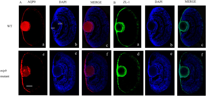
Figure Caption
Fig. 4 Distribution of MIP and ZL-1 protein in mutant lens (A) While mutant lens demonstrated a great decline in Mip protein (AQP0) expression compared to that in WT lens. (B) Diffuse distribution of ZL-1 positive signal was observed in the mutant lens while it was mainly located in the lens epithelial cells and elongating fiber cells in WT fish lens. AQP0: red, ZL-1: green; DAPI: blue. Scale bar = 25 μm.
Figure Data
Acknowledgments
This image is the copyrighted work of the attributed author or publisher, and
ZFIN has permission only to display this image to its users.
Additional permissions should be obtained from the applicable author or publisher of the image.
Reprinted from Gene expression patterns : GEP, 49, He, M., Zhou, G., Lin, Q., Zhou, N., The role of mip in the development of lens in zebrafish, 119330, Copyright (2023) with permission from Elsevier. Full text @ Gene Expr. Patterns