
- Title
-
Embryonic and aglomerular kidney development in the bay pipefish, Syngnathus leptorhynchus
- Authors
- Maters, B.R., Stevenson, E., Vize, P.D.
- Source
- Full text @ PLoS One

A. Blastula/disc stage. The black arrow indicates the lateral border of the dense disc of cells. The white arrow indicates a larger disc of more dispersed material that the cellular disc sites upon. Approximate equivalent; medaka stage 11–12. B. Early segmentation stage. Anterior end of embryo is to the left. Approximate medaka stage 18–19. C. Late segmentation stage, lateral view. Retina obvious. Approximate medaka stage equivalent is stage 22. D. Early pharyngula stage. Retina obvious, extensive somitogenesis. Posterior tail is not attached to yolk sac. Approximate equivalent; medaka stage 25, except the tail is longer and detached in the pipefish. E. Mid-pharyngula stage. No snout, no fin blastemas. F. Mid pharyngula stage II. Slight protuberance in future snout region (white arrow). Dorsal and caudal fin blastemas present, first dorsal fin rays are emerging, no rays in caudal fin. G. Late pharyngula stage. Future snout small. Dorsal fin rays still forming, first rays in caudal fin are apparent. H. Late pharyngula stage. Future snout small. Dorsal fin rays still forming, first rays in caudal fin are apparent. I. Early hatching stage. Snout distinct, jaw visible and mouth can open. Dorsal and caudal fin rays are well developed. Yolk sac is 50% absorbed. J. Mid hatching stage. Snout becoming longer, tail extending, yolk sac remnant rapidly decreasing in size, approximately 25% of original size. K. Late hatching stage, early fry stage, shortly before emergence. Elongated snout, well developed dorsal fin. Yolk sac has been resorbed. Birth imminent. The panel in K illustrates the anal fin in this embryo under higher magnification. Further details on the developing snout and fins are present in the following figure. |

Scale bars in A, C, E and F represent 100 microns, white arrows indicate fin rays. A. Mid-pharyngula stage, no mouth, dorsal fin and caudal fin blastemas. Otolith visible. B. Similar to A, but slightly older. C. Mid pharyngula stage. Slight protuberance in future mouth region. Dorsal fin rays are emerging, no rays in caudal fin. D. Late pharyngula stage. Future snout small. Dorsal fin rays still forming, first rays in caudal fin are apparent. E. Early hatching stage. Snout distinct, jaw visible and mouth can open. Dorsal and caudal fin rays are well developed. Yolk sac is 50% absorbed. Melanocytes star shaped. F. Late hatching stage, early fry stage, shortly before emergence. Elongated snout, well developed dorsal fin. Yolk sac has been resorbed and birth is imminent. |

A. Transverse section of a JB4 plastic embedded specimen. Various dorsal tissues surrounding the kidney tubules are labelled. B. Enlargement of panel A. The red arrows indicate the kidney tubules. |

Amino acid residues conserved in all species are indicated with a ‘*’, highly similar amino acids with a ‘:’ and similar amino acids with a ‘.’. The NCBI accession numbers of aligned proteins are; black sea bream, ABR10300.1, tarwhine, AAT48993.1; zebrafish AAI63629.1; tetraodon, CAF97804.1. The first conserved amino acid of the pipefish protein, the lysine residue (K), corresponds to amino acid 25 of the zebrafish reference protein. |
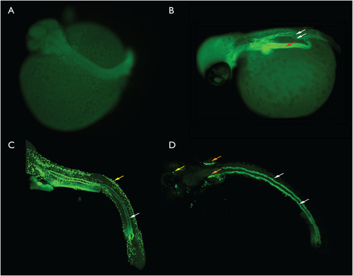
A. Late segmentation stage, no kidney is visible. B. Early pharyngula, paired kidney tubules are present and atp1a positive (white arrows). The gut tube is also fluorescent (red arrow). C. Scanning confocal generated image stack illustrating the kidney tubules (white arrow) and ionocytes (yellow arrow) in a late pharyngula embryo. D. Late pharyngula scanning confocal generated image stack. This stack was collected only in the plane of the kidney so most other labelled structures were not present. An unknown structure (orange arrows), possibly the developing gills and some ionocytes (yellow arrow) were also present in the image stack. For the purpose of scale, the eye in the early pharyngula in panel 5B is approximately 150 microns in diameter. |

A-C, three views of a single set of aglomerular tubules. A. Dorsal view, anterior left, confocal stack. B. AMIRA generated surface model of the data illustrated in panel A. C. The same model as illustrated in panel B rotated so that the anterior portion of the kidney is on the lower right, closest to the viewer. a, anterior; p, posterior. D-F, three views of a single set of aglmoerular tubules. D. Confocal stack. E. AMIRA surface model of the same set of tubules as illustrated in D. F. The model illustrated in E rotated with the posterior end now closest to the viewer. a, anterior; p, posterior. 3D rotations of these models can be viewed in |