
|
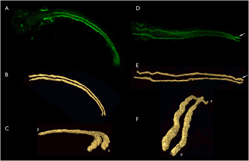
Fig 6
A-C, three views of a single set of aglomerular tubules. A. Dorsal view, anterior left, confocal stack. B. AMIRA generated surface model of the data illustrated in panel A. C. The same model as illustrated in panel B rotated so that the anterior portion of the kidney is on the lower right, closest to the viewer. a, anterior; p, posterior. D-F, three views of a single set of aglmoerular tubules. D. Confocal stack. E. AMIRA surface model of the same set of tubules as illustrated in D. F. The model illustrated in E rotated with the posterior end now closest to the viewer. a, anterior; p, posterior. 3D rotations of these models can be viewed in