
|
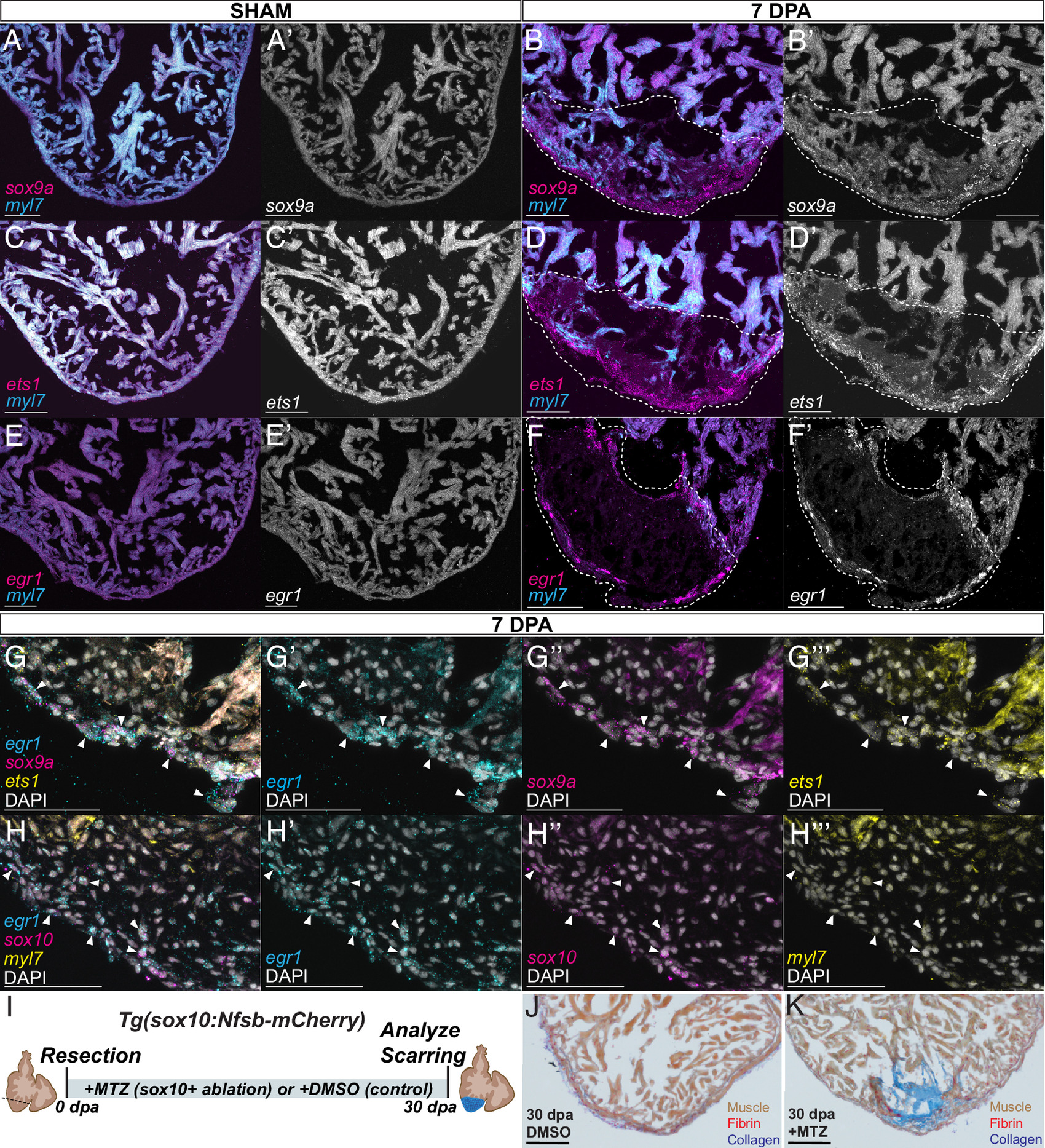
Fig. 4 A developmental CdNC subcircuit is expressed during adult cardiac regeneration. HCR staining of sham and injured 7dpa ABWT hearts, respectively, for myl7 and (A and B) sox9a (n = 12/12), (C and D) ets1 (n = 11/11), and (E and F) egr1 (n = 15/15). (A′–F′) Grayscale of respective sox9a, ets1, and egr1 stainings. (Scale bar, 100 μm.) (G) HCR costaining on ABWT 7dpa hearts for sox9a, egr1, ets1, and DAPI (n = 8/8). Arrows indicate some of the cells positive for all three probes. Single channel images, with DAPI, of, sox9a, egr1, and ets1 are shown respectively in (G′ and G″′). (Scale bar, 50 μm.) (H) HCR costaining on ABWT 7dpa hearts for sox10, egr1, myl7, and DAPI (n = 10/11). Arrows indicate a subset of the cells double-positive for sox10 and egr1 probes. Single channel images, with DAPI, of egr1, sox10, and myl7 are shown respectively in (H′ and H″). (I) Diagram of the sox10+ ablation scheme. Tg(sox10:Nfsb-mCherry) injured fish are maintained in MTZ for the duration of the regeneration time course and hearts are harvested at 30 dpa. (J) Injured hearts maintained in DMSO to 30 dpa retain little scarring (n = 3/3), whereas (K) injured hearts maintained in MTZ, ablating sox10+ cells, maintain a scar to 30 dpa (n = 5/5). (Scale bar, 100 μm.)