
|
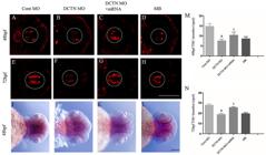
Fig. 1 Knockdown of the dynactin gene led to a decrease of dopaminergic neurons in the diencephalon. Immunofluorescence was used to observe the changes of tyrosine hydroxylase (TH)-positive neurons in the diencephalon of zebrafish at 48 and 72 hpf. A and E, Control MO group; B and F, DCTN MO group; C and G, DCTN MO+mRNA group; D and H, MB group. Scale bar, 100 μm. Expression of TH in the diencephalon of zebrafish was detected at 48 hpf by in situ hybridization. I, Control MO group; J, DCTN MO group; K, DCTN MO+mRNA group; L, MB group. The white and black dotted lines indicate the ventral area of the zebrafish diencephalon. Scale bar, 200 μm. M and N, Number of TH-positive neurons in zebrafish diencephalon at 48 and 72 hpf. Data are reported as means±SD. aP<0.0001, compared with the Cont MO group; bP<0.05, compared with the DCTN MO group (two-way ANOVA, Tukey post hoc, n=15-30 zebrafish per group). Cont MO: control morpholino; DCTN MO: dynactin morpholino; DCTN MO+mRNA: dynactin morpholino+dynactin mRNA; MB: mycalolide B.