IMAGE

Fig. 4.

ID
ZDB-IMAGE-250422-104
Source
Figures for Laverde et al., 2025
Image
Figure Caption

Fig. 4.

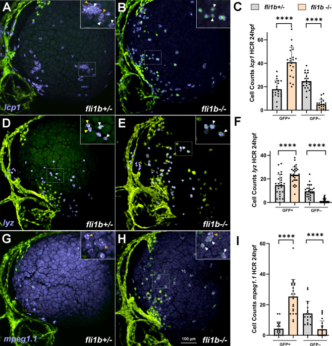
Myeloid marker expression analysis at 24 hpf. HCR analysis for expression of the pan-myeloid marker lcp1 (A-C), the neutrophil marker lyz (D-F) and the macrophage marker mpeg1.1 (G-I) in fli1b+/− control and fli1b−/− mutant embryos. Two populations of lcp1+ GFP+ (or lyz+ GFP+ or mpeg1.1+ GFP+) and lcp1+ GFP (or lyz+ GFP or mpeg1.1+ GFP) were observed. Boxed areas are shown at a higher magnification in the upper right inserts. White arrowheads show GFP+ myeloid cells, whereas yellow arrowheads show GFP myeloid cells. Only myeloid cells located at the yolk surface were quantified. Note that the number of GFP+ myeloid cells was significantly increased in fli1b−/− embryos, whereas the number of GFP myeloid cells was reduced. Myeloid cell GFP expression was faint in fli1b+/− embryos and much brighter in fli1b−/− embryos; both weak and strong GFP+ cells were included in GFP cell counts. Bars show mean±s.d. ****P<0.0001; Student's two-tailed unpaired t-test.

Acknowledgments
This image is the copyrighted work of the attributed author or publisher, and ZFIN has permission only to display this image to its users. Additional permissions should be obtained from the applicable author or publisher of the image. Full text @ Biol. Open