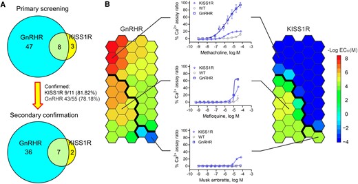
Fig. 3 Secondary Ca2+ flux assay confirmation. A, The Venn diagram shows the secondary Ca2+ flux assay confirmation results. B, The heat map shows the half maximum effective value (EC50) distribution of all 45 compounds identified and confirmed from secondary assays based on the self-organizing map (SOM) algorithm. The left panel shows the data from the HEK293-GnRHR cell line, and the right panel shows the data from the HEK293-KISS1R cell line. Red indicates a compound with a low EC50 and high potency, and blue indicates a compound with a high EC50 and low potency. Dark blue indicates a compound was inactive in the tested cell line. Compounds can be separated into 3 groups based on their EC50 in each cell line through the SOM algorithm, as indicated by the bold black line. A representative compound for each group and its concentration-response curve are shown in the middle panel.
Image
Figure Caption
Acknowledgments
This image is the copyrighted work of the attributed author or publisher, and
ZFIN has permission only to display this image to its users.
Additional permissions should be obtained from the applicable author or publisher of the image.
Full text @ Endocrinology

