
|
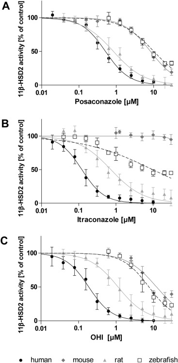
Fig. 1 Inhibition of human, mouse, rat and zebrafish 11β-HSD2 activity. Lysates of HEK-293 cells expressing recombinant 11β-HSD2 of the respective species were incubated for 10 min (human) or 20 min (mouse, rat, zebrafish) in the presence of 50 nM cortisol, 500 μM NAD+ and increasing concentrations of posaconazole (A), itraconazole (B) or OHI (C). Substrate conversion was normalized to that of the vehicle control (DMSO; 0.3% for posaconazole, 0.6% for itraconazole and 1.2% for OHI). Inhibition curves were fitted and analyzed by non-linear regression. Data shown are from at least three independent experiments and represent mean ± SD.