
Fig. 2
- ID
- ZDB-FIG-250820-2
- Publication
- Petrova et al., 2025 - Semaphorin 3F is elevated in serum of heart failure patients and inhibits cardiac angiogenesis via the VEGF/Akt/eNOS pathway
- Other Figures
- All Figure Page
- Back to All Figure Page
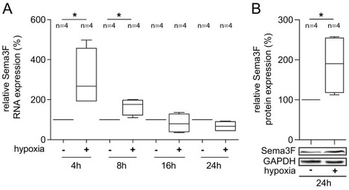
Sema3F expression is regulated by hypoxia in cardiac endothelial cells. (A) The expression of Sema3F RNA was increased in endothelial cells (HCECs) under hypoxic conditions, with the highest levels being observed after 4 h. HCECs were exposed to hypoxia (1 % O2) for the indicated times, after which the cells were harvested, RNA was isolated and reverse transcribed. cDNA was then used for the quantification of Sema3F expression by qRT-PCR. The data presented here is derived from four independent experiments (biological replicates, n = 4), and the statistical analysis employed was the Man-Whitney test. A p-value <0.05 is considered to be significant and marked with an asterisk (*). (B) Sema3F protein was significantly increased in response to 24 h of hypoxia in comparison to normoxia. For Western blot analysis HCECs were lysed and Sema3F protein levels were detected by an anti-Sema3F antibody. GAPDH was used as a loading control. Representative blots from four independent experiments are presented (biological replicates, n = 4). The results were analyzed using the Mann-Whitney test (*p < 0.05). |