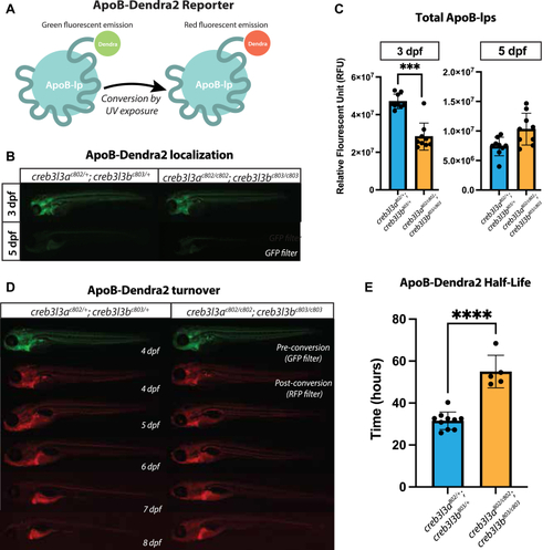
ApoB-Dendra2 reporter localization in creb3l3 mutants. A: Schematic of ApoB-Dendra2 fusion. Dendra2 is endogenously fused to the C terminus of ApoBb.1 for in vivo characterization of the ApoB-lp profile. The unconverted form of Dendra2 is visualized with a GFP filter. UV exposure irreversibly photoconverts Dendra2 to emit fluorescence visualized by the RFP filter. B: GFP filter showing ApoB-Dendra2 signal in 3 and 5 dpf zebrafish in creb3l3 double homozygous and double heterozygous siblings. Images are representative of 3 independent clutches; in each clutch, there were 4–10 animals per genotype. All fish used in these experiments were homozygous for the ApoBb.1-Dendra2 reporter and were compared to apoBb.1+/+ for background fluorescence. Unpaired t-tests ∗∗∗P < 0.0005. C: Quantification of fluorescent signal at 3 and 5 dpf. n = 9–10 animals per genotype. D: Representative images of ApoB-Dendra2 reporter in creb3l3 double homozygous mutants and creb3l3 double heterozygous siblings. The same fish is imaged every 24 h from 4 to 8 dpf. Before UV exposure (preconversion) images were taken using the GFP filter and pseudo-colored green. After UV exposure (post conversion) images were taken using an RFP filter and pseudo-colored red. Images are representative of three independent clutches; in each clutch, there were 4–10 animals per genotype. E: Quantification of RFP fluorescent signal from 4 to 8 dpf to measure half-life of ApoB-Dendra2 particles. N = 3 clutches; n = 4–10 individuals per genotype per clutch; unpaired t-tests ∗∗∗∗P < 0.0001. ApoB-lp, apolipoprotein-B–containing lipoprotein; Creb3l3, cAMP-responsive element-binding protein 3–like 3; dpf, days post fertilization.
|
