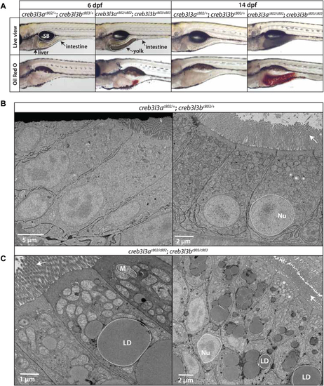
Accumulation of cytoplasmic lipid droplets in the enterocyte of creb3l3 mutant animals. A: Representative brightfield live images of creb3l3ac802/+;creb3l3bc803/+ and creb3l3ac802/c802;creb3l3bc803/c803 zebrafish larvae at 6 dpf. Unfed 6 dpf creb3l3 double mutants retain yolk and exhibit a yolk opacity phenotype compared to double heterozygous siblings, which suggests abnormal lipid droplet accumulation in the yolk syncytial layer. creb3l3ac802/c802;creb3l3bc803/c803 animals exhibit abnormal lipid accumulation by Oil Red O (ORO) staining in the posterior region of the yolk at 6 dpf. Images are representative of 3 independent experiments with 5–9 animals per experiment. Fasted 14 dpf creb3l3 mutants have opaque intestines under brightfield (Live view) and increased ORO staining throughout the intestine in fasted fish at 14 dpf. creb3l3ac802/+;creb3l3bc803/+ animals do not stain with ORO. Images are representative of 3 independent experiments with 5–9 animals per experiment. B: Representative electron microscopy images of creb3l3ac802/+;creb3l3c803/+ and creb3l3ac802/c802;creb3l3bc803/c803 animals fasted at 14 dpf (C). Large lipid droplets (LD) are present in creb3l3ac802/c802;creb3l3bc803/c803 animals. Images are representative of 3–6 animals per genotype. The nucleus and mitochondria are outlined by white dotted lines and lipid droplets are outlined by a solid line. Arrows points to microvilli on the apical side of the enterocyte. Creb3l3, cAMP-responsive element-binding protein 3–like 3; dpf, days post fertilization; Nu, nucleus; M, mitochondria; SB, swim bladder.
|
