
Fig. 2
- ID
- ZDB-FIG-250207-13
- Publication
- Kasuya et al., 2024 - Identification of KCNE6, a new member of the KCNE family of potassium channel auxiliary subunits
- Other Figures
- All Figure Page
- Back to All Figure Page
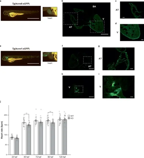
Functional characterizations of KCNE6 in zebrafish.a Representative images of the Tg(kcne6:eGFP) line at 72 hpf under eGFP fluorescence. The scale bar indicates 0.5 mm. The inset shows a close-up view of the heart. b–d Immunofluorescence staining of a heart section from the adult Tg(kcne6:eGFP) line using the anti-GFP antibody. The scale bars indicate 0.5 mm in (b) and 0.1 mm in (c, d). AT atrial myocyte. V ventricular myocyte. BA bulbus arteriosus. e Representative images of the Tg(kcne1:eGFP) line at 72 hpf under eGFP fluorescence. The scale bar indicates 0.5 mm. The inset shows a close-up view of the heart. f–i Immunofluorescence staining of a heart section from the adult Tg(kcne1:eGFP) line using the anti-GFP antibody. The scale bars indicate 0.5 mm in (f, h) and 0.1 mm in (g, i). j Heart rates in the WT and kcne6 KO lines. In each line, ten healthy embryos from three independent pairs (n = 30 in total) were collected at 24 hpf and monitored from 24 hpf to 120 hpf. Student’s t-test was used to compare the WT and kcne6 KO lines and significance was assigned at p < 0.05 (*p < 0.05, **p < 0.01, and ***p < 0.001). The exact p-values were 0.0013 at 48 hpf and 0.0087 at 96 hpf. |