
Fig. 3
- ID
- ZDB-FIG-250128-54
- Publication
- Gimenez et al., 2024 - Cohesin rad21 mutation dysregulates erythropoiesis and granulopoiesis output within the whole kidney marrow of adult zebrafish
- Other Figures
- All Figure Page
- Back to All Figure Page
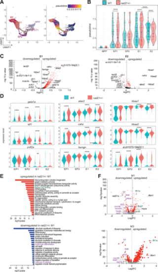
Erythroid differentiation program is altered in rad21+/− WKM. A: UMAP of scRNA-seq data showing erythroid cell clusters and Monocle 3 pseudotime trajectory within the erythroid lineage starting from MPP. B: violin plots showing the distribution of the pseudotime values for MPP and erythroid cells. Indicated in blue dots are the median pseudotime values for each cluster. Significance was determined using the Wilcoxon rank-sum test. ****P < 0.0001. C: volcano plot showing DEGs identified from comparison of rad21+/− versus WT in E1 and E2. Red dot signifies statistically significant (P < 0.05) DEGs. D: violin plots showing the expression of erythroid associated genes. Significance is from the scRNA-seq DESeq2 Wald test. **P < 0.01, ***P < 0.001, ****P < 0.0001 (Supplemental Table S3). E: GO enrichment (using Metascape) on the significant DEGs (P ≤ 0.05) identified in M1 from comparison of rad21+/− versus WT cells. Complete list of pathways and the associated DEGs are provided in Supplemental Table S5. F: volcano plot showing DEGs identified in M1 and M3 in comparison of rad21+/− versus WT. Red dot signifies statistically significant (adjusted P ≤ 0.05) DEGs. Shown in green are iron storage genes and in purple are the erythrocyte and cytokine genes. WKM, whole kidney marrow; WT, wild type. |