
Fig. 1
- ID
- ZDB-FIG-250103-18
- Publication
- Kong et al., 2024 - Apigenin attenuates cisplatin-induced hair cell damage in the zebrafish lateral line
- Other Figures
- All Figure Page
- Back to All Figure Page
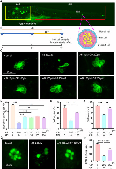
Apigenin protects against cisplatin-induced ototoxicity. (A) Confocal image of a 5 dpf Tg (Brn3c:mGFP) zebrafish larva. The green fluorescent point circled by the yellow rectangle represents the anterior lateral line system (aLL), while the green fluorescent point circled by the red rectangle represents the posterior lateral line system (pLL). A schematic lateral view of a 5 dpf neuromast illustrating different cell types is shown below. (B) Schematic diagram illustrating the experimental workflow of zebrafish. (C) Live confocal imaging of Tg (Brn3c:mGFP) HCs (green) in L1 neuromast from larvae at 5 dpf. Zebrafish were exposed to varying concentrations of cisplatin with and without apigenin. Control animals received the vehicle alone (DMSO). (D) Quantification of the number of HCs in L1 neuromast after different treatments, represented as mean ± SEM (n = 10). (E) Peak velocity and (F) swimming distance of 5 dpf zebrafish larvae, indicative of auditory function, assessed by startle response. (G) Staining of hair cell damage with 2-(4-[dimethylamino]styryl)-N-ethylpyridinium iodide (DASPEI) showed that treatment with cisplatin decreased the number of hair cells in L1 neuromast. However, the co-treatment with 100 μM apigenin protected reducing number of hair cell staining with DASPEI. (H) Average DASPEI stained area in L1 neuromast in different groups were shown NM: neuromast; API: apigenin; CP: cisplatin. Statistical significance indicated as ∗, ∗∗, ∗∗∗and ∗∗∗∗ above the bars (∗∗∗∗P < 0.0001, ∗∗∗P < 0.001, ∗∗P < 0.01, ∗P < 0.05). Scale bar equals 25 μm. (For interpretation of the references to colour in this figure legend, the reader is referred to the Web version of this article.) |