
Fig. 7
- ID
- ZDB-FIG-240223-23
- Publication
- Thomasen et al., 2023 - SorCS2 binds progranulin to regulate motor neuron development
- Other Figures
- All Figure Page
- Back to All Figure Page
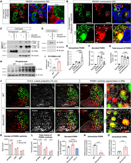
SorC2 binds PGRN to control its trafficking and secretion (A and B) IF of HEK293 cells overexpressing hPGRN and hSorCS2 as co-culture (trans configuration) (A) or co-expressing the plasmids in the same cell (cis configuration) (B). hPGRN and hSorCS2 do not co-localize in trans (A), but they do in vesicular structures and plasma membrane when in cis (arrows) (B). Hoechst-labeled nuclei (blue). (C) Co-IP of transfected HEK293 cells. SorCS2 binds PGRN in cis but not in trans. Arrows point at SorCS2 isoforms with the proform binding hPGRN. β-Actin served as a loading control for the input and negative control for the co-IP. (D) Immunoblotting of secreted hPGRN in transfected HEK293 cells. (E‒G) Compiled quantification of independent input samples from lysates and cell media blotted for hPGRN and HA-hPGRN. n = 4/5. When in cis, SorCS2 retains hPGRN intracellularly (E) while reducing PGRN secretion (F). (G) Total PGRN levels are decreased in cis but not in trans configuration. (H and I) (H) Immunoblotting of spinal cord from P3 pups (n = 4 pups/genotype). (I) The band intensity is normalized to the loading control. (J‒L) Confocal scanning and analysis of PGRN+ fluorescent signal in posterior coronal sections of brachial MNs at E12.5. n = 6 embryos/genotype. (J) IF staining of PGRN (left panels) was segmented in Imaris (right panels) as spots (gray). (K and L) LMC and MMC populations were either pooled (“all MNs”) or distinguished by Islet1 and Foxp1 IF. (K) Quantification of PGRN+ particle count. (L) Quantification of total volume of PGRN+ particles. (M) ELISA of conditioned media from primary cortical neuron cultures (DIV 7). Grn+/− served as semi-negative control. n = 3. (N and O) Immunoblot quantifications of lysates from the cortical cultures (n = 4). All graphs shown mean ± SEM. See also Figures S11 and S12; Video S10. |