
FIGURE
Fig. S4
Fig. S4
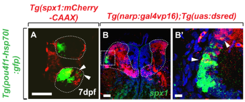
Expression of spx1 in dorsal Hb. (A) Dorsal view of the Hb region in Tg(spx1:mCherry-CAAX);Tg(pou4fl-hsp70l:gfp) larva at 7 dpf. Arrowheads indicate the spx1 and pou4fl+ cells in Hb. (B, B’) Transverse section of the Hb in Tg(narp:gal4vp16);Tg(uas:dsred) adult brain labelled by spx1 RNA and dorsal is to the top. (B’) is a high magnification image of the box area in (B). Arrowhead indicates the spx1 and narp+ cells in lateral division of the dorsal Hb. Abbreviation: Hb, habenula; IPN, interpeduncular nucleus. Scale bar: 50 μm in A and B. 10 μm in B’. |
Expression Data
Expression Detail
Antibody Labeling
Phenotype Data
Phenotype Detail
Acknowledgments
This image is the copyrighted work of the attributed author or publisher, and
ZFIN has permission only to display this image to its users.
Additional permissions should be obtained from the applicable author or publisher of the image.
Full text @ Sci. Rep.