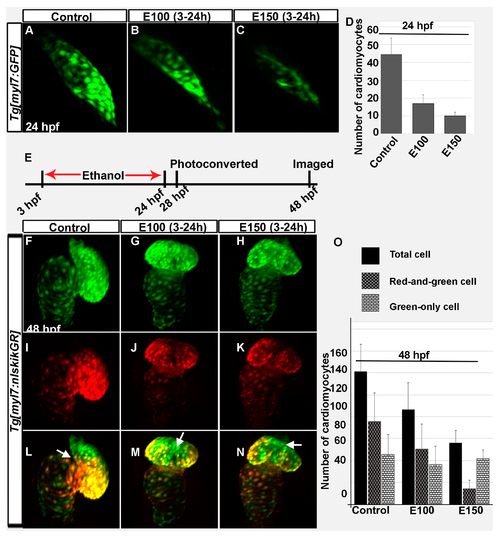
Ethanol exposure during embryogenesis reduced the number of early- and late-added cardiomyocytes in the heart. (A–C) 3D renderings of confocal sections of Tg(myl7:GFP) embryos showed GFP positive FHF derived cardiomyocytes in the linear heart tube in the control (A) and ethanol-exposed embryos (B,C); (D) Graph shows reduced number of FHF derived cardiomyocytes in ethanol-exposed embryos at 24 hpf. p < 0.001; (E) Schematic shows the time of ethanol treatment, photoconversion, and image acquisition in this experiment; (F–N) 3D renderings of confocal sections of photoconverted Tg(myl7:nlskikGR) embryos showed hearts in the control and ethanol-treated embryos. Note the green-only cardiomyocytes (cardiomyocytes added after photoconversion) in the anterior region of the ventricle in control embryos (A,L); but in the mid-ventricular region in ethanol-treated embryos (G–N); White arrows: green-only cells; (O) Graph shows the quantification of total, red-and-green (early-added cardiomyocytes) and green-only (late-added cardiomyocytes) cardiomyocytes at 48 hpf.
|
