|
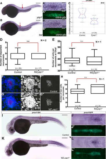
yap1 and taz are ubiquitously expressed at 30 hpf. Zyap1/ mutant embryos have fewer cells compared to sibling controls. (A–B′′) 30 hpf cldnb:gfp embryos stained with a yap1 (A–A′′) or a taz (B–B′′) ISH probe and an anti-GFP antibody (A′′, B′′). (C–E, H) Boxplot showing pLLP cell counts (C), number of deposited neuromasts (D), cell counts (E), and number of hair cells (H) per neuromast in the indicated experimental conditions. Cell counts were performed in the neuromast closest to the end of yolk extension (n3 or n4) at 72 hpf in (E–H). (F–G′′) MIP of Z-stacks of a deposited neuromast (n3 or n4) stained with an anti-HCS1 antibody (hair cells, red) and DAPI (cell nuclei, blue). (I–L′) ISH with a prox1 probe and an anti-GFP antibody staining (J′, L′) on embryos with the indicated genetic background. Red arrows point to the pLLP (A, B, I and K) (Figure 5—source data 3, 4).
|