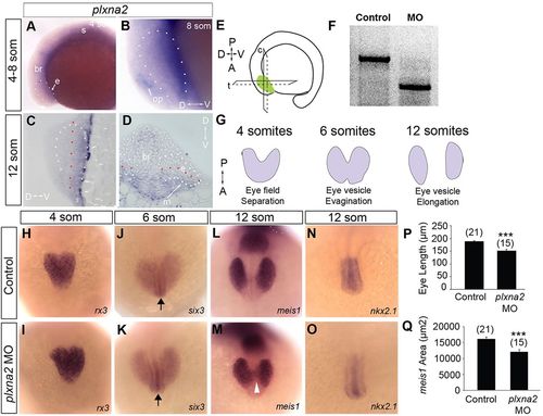
plxna2 morphants exhibit small eye vesicles. (A-D) RNA in situ hybridization for plxna2 viewed in lateral wholemounts (A,B), and in sagittal (C) and transverse (D) sections. At 4-12 somites, plxna2 mRNA is in the ventral, but not dorsal, eye (e) vesicle (outlined in white), as well as in the mesenchyme (m), somites (s) and brain (br). op, olfactory placode. Red dotted line separates the eye vesicle into approximate dorsal and ventral domains. (E) Transverse (t) and coronal (c) planes of section. (F) RT-PCR indicates the expected mis-splicing of plxna2 mRNA in the morphants. (G) A dorsal view of the events of eye vesicle formation. (H-O) Dorsal views of whole-mount RNA in situ hybridization of control (H,J,L,N) and plxna2 morphant (I,K,M,O) embryos. rx3 (H,I), six3 (J,K) and meis1 (L,M) labeling. Arrows indicate the hypothalamus; the arrowhead marks meis1+ cells that bridge the eye vesicles in the plxna2 morphant. (N,O) nkx2.1 labeling of hypothalamus. (P,Q) Quantitation of the length (P) and area (Q) of the meis1+ domain. n=2. Error bars indicate s.e.m. ***P<0.001 unpaired Student′s t-test.
|
