
FIGURE
Fig. S1
- ID
- ZDB-FIG-120824-15
- Publication
- Chen et al., 2012 - Haemodynamics-driven developmental pruning of brain vasculature in zebrafish
- Other Figures
- All Figure Page
- Back to All Figure Page
Fig. S1
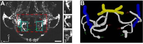
Midbrain vasculature at 1.5 dpf. (A) Projected confocal image of a 1.5-dpf Tg(kdrl:eGFP) larva showing that some angiogenic sprouts (green arrows) were observed in the midbrain. Inset 1, filopodium-like sprout; Insets 2 and 3, sprouts with an expanded tip. Red and green arrows point to sprouts originated from the choroidal vascular plexus (CVP) or midbrain vasculature, respectively. The dashed square delineates the midbrain position. Scale, 50 μm. (B) 3-D reconstruction of the midbrain vasculature shown in (A). Yellow, basal communicating artery (BCA); white, midbrain vessels; blue, CVP. |
Expression Data
Expression Detail
Antibody Labeling
Phenotype Data
Phenotype Detail
Acknowledgments
This image is the copyrighted work of the attributed author or publisher, and
ZFIN has permission only to display this image to its users.
Additional permissions should be obtained from the applicable author or publisher of the image.
Full text @ PLoS Biol.