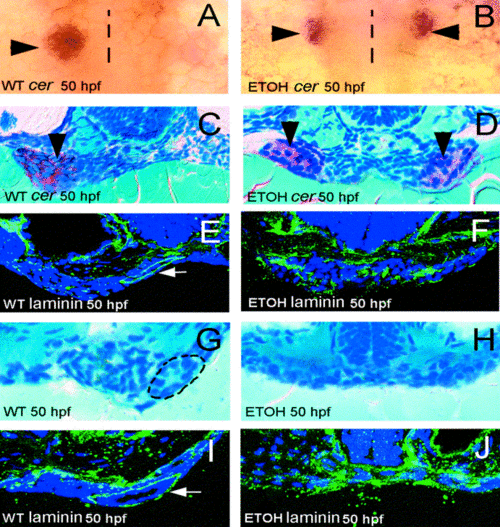
The zebrafish liver and pancreas form independently of the gut tube. (A, B) Whole-mount RNA in situ hybridization using a ceruloplasmin (cer) probe, dorsal view. Compared with WT (A), liver duplication (arrows) is seen in 50-hpf ethanol-treated embryos (B). (C, D) Histological cross-sections through the liver of 50-hpf WT (C) and ethanol-treated (D) embryos processed for cer in situs. Anterior endoderm spans the midline between the duplicated livers (arrows in D) of ethanol-treated embryos. (E, F) Laminin immunostainings from a comparable region of 50-hpf WT (E) and ethanol-treated (F) embryos. Organized endoderm present within the developing esophagus (white arrow) of 50-hpf WT embryos is not identifiable in ethanol-treated embryos (F). (G) Histological cross-sections through the rostral gut of 50-hpf WT embryo. (H) Endoderm in a comparable region of 50-hpf ethanol-treated embryo spans the midline and is unorganized. (I, J) Immunoreactive laminin in a WT 50-hpf embyro (I) encircles the gut tube, but is widely dispersed in unorganized endoderm of ethanol-treated embryos (J). Dotted line in (A, B) represents the embryonic midline. Circle in (G) surrounds the gut tube.
|
