
FIGURE
Fig. S4
Fig. S4
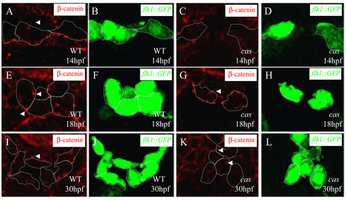
Adherens junction formation during vascular development. Transverse sections of wild-type embryos (A,B,E,F,I, J) and endoderm-less embryos [Tg(flk1:EGFP)s843;cas mutants] (C,D,G,H,K,L) visualized for &b;-catenin (red) (A,C,E,G,I,K) and GFP (green) (B,D,F,H,J,L). The sections are at the level of the 6th (A-H) and 12th somite (I-L). White arrowheads indicate adherens junctions between endothelial cells, which appear after the cells reach the midline. |
Expression Data
Expression Detail
Antibody Labeling
Phenotype Data
Phenotype Detail
Acknowledgments
This image is the copyrighted work of the attributed author or publisher, and
ZFIN has permission only to display this image to its users.
Additional permissions should be obtained from the applicable author or publisher of the image.
Full text @ Development