
- Title
-
Pax1a-EphrinB2a pathway in the first pharyngeal pouch controls hyomandibular plate formation by promoting chondrocyte formation in zebrafish
- Authors
- Jeon, H., Jin, S., Kim, J., Joo, S., Choe, C.P.
- Source
- Full text @ Front Cell Dev Biol
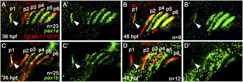
Expression of |

Requirement of |
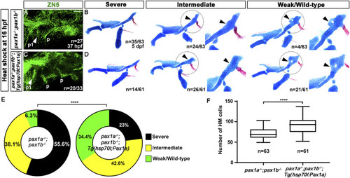
Rescue of the defects in the first pouch and hyomandibular cartilage in |
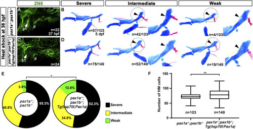
Rescue of the defects in the hyomandibular cartilage in |

Loss of skeletogenic mesenchyme in the anterior region of developing hyomandibular plate in |
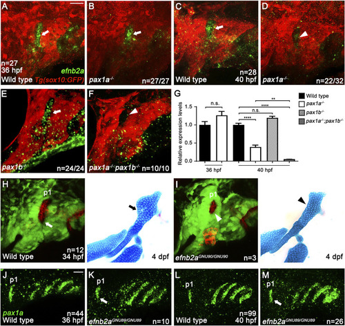
Genetic linkage of EphrinB2a with Pax1a in hyomandibular plate development. |

Requirement of Pax1a-EphrinB2a in the first pouch for hyomandibular plate development. |

Requirement of Pax1a-EphrinB2a after pouch formation in hyomandibular plate development. |
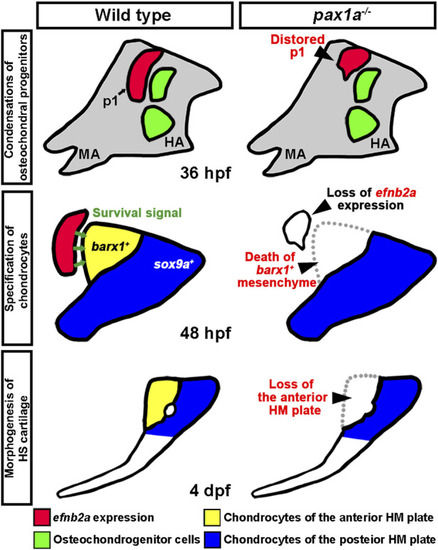
A model for the role of Pax1a in hyomandibular plate development. In wild types, Pax1a is necessary for the morphogenesis of the first pouch that forms at 18 hpf, with the loss of Pax1a leading to a distorted first pouch. After pouch formation, |