
- Title
-
Zebrafish-Encoded 3-O-Sulfotransferase-3 Isoform Mediates Herpes Simplex Virus Type 1 Entry and Spread
- Authors
- Hubbard, S., Darmani, N.A., Thrush, G.R., Dey, D., Burnham, L., Thompson, J.M., Jones, K., and Tiwari, V.
- Source
- Full text @ Zebrafish

Expression of zebrafish (ZF) 3-O-sulfotransferase-3 (3-OST-3) isoform in wild-type Chinese hamster ovary (CHO-K1) cells results in herpes simplex virus type 1 (HSV- 1) entry. (A) Dose–response curve of HSV-1 entry into CHO-K1 cells expressing ZF 3-OST-3 isoform. Resistant wild-type CHO-K1 cells were transfected with ZF 3-OST-3 at 2.5 μg DNA, which in resulted HSV-1 KOS (gL86) entry, similar to human 3-OST-3 (H 3-OST-3) expression. Cells transfected with empty vector pDream2.1 at 2.5 μg DNA was used as a negative control. The enzymatic activity was measured at an optical density of 410nm (OD410). Each value shown is the mean of three or more determinations (±standard deviation). (B) HSV-1 entry into ZF 3-OST-3 isoform expressing CHO-K1 cells was confirmed by X-gal staining. CHO-K1 cells (4 x 10⁶ cells) expressing H 3-OST-3 (panel a) and ZF 3-OST-3 (panel b) were challenged with β-galactosidase-expressing recombinant HSV-1 KOS (gL86) at 25 pfu/cell. Wild-type CHO-K1 cells expressing pDream2.1 (panel c) were also infected in parallel as a negative control. After 6 h of infection at 37°C, cells were washed with phosphate-buffered saline, fixed, permeabilized, and incubated with X-gal (5 bromo-4 chloro-3-indoyl-β-D-galactosidase) at 1.0mg/mL, which yields an insoluble blue product upon hydrolysis by β-galactosidase. Blue cells (representing viral entry) were identified. Microscopy was performed using a 20x objective of Nikon D-Eclipse-C1. (C) Observation of HSV-1 entry in cultured CHO-K1 expressing ZF 3-OST-3 isoform. Confluent monolayers of ZF 3OST-3 CHO-K1 cells (panel b) were infected with green fluorescent protein–tagged capsid of HSV- 1 (VP26-green fluorescent protein) at 25 pfu/cell for 90 min. In parallel CHO-K1 cells expressing pDream2.1 (panel c) and H 3-OST-3 (panel a) were used as negative and positive controls, respectively. At 90 min postinfection, cells were fixed with 2% formaldehyde and 0.2% glutaraldehyde, and stained with 10nm rhodamine-conjugated phalloidin (red) for F-actin. The images were taken using a fluorescent confocal microscope (Nikon D-Eclipse-C1) at 40x objective. |
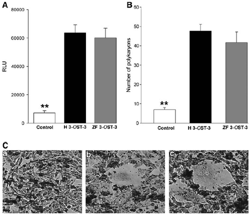
(A) ZF 3-OST-3-expressing target CHO-K1 cells gain the ability to fuse with effector cells coexpressing HSV-1 glycoproteins gB, gD, gH, and gL. The target cells were transfected with plasmids expressing H 3-OST-3 and ZF 3-OST-3 (as indicated), and luciferase reporter gene. The effector cells were transfected with HSV-1 glycoproteins gB, gD, gH, and gL, and T7 RNA polymerase. A luciferase reporter assay was performed 24 h after the two cell populations were mixed together. Cell fusion was measured in relative luciferase units (RLUs) using a Sirius luminometer (Berthold Detection System). (A) Expression of ZF 3-OST-3 isoform results in the fusion of CHO-K1 cells with HSV-1-glycoprotein-expressing cells as measured by a luciferase assay system with reporter lysis buffer (Promega). The data shown are the means of triplicate measures and are representative of three independent experiments. Double asterisks indicate significant difference from other treatments ( p < 0.01, t-test); error bars represent SD. (B) Quantitative determination of the number of fused polykaryocytes formed after coculture of target cells expressing either ZF-encoded 3-OST-3 or human-encoded 3-OST-3 receptors with the effector CHO-K1 cells expressing HSV-1 glycoproteins. Target CHO-K1 cells expressing empty vector (pDream2.1) cultured with effector CHO-K1 cells were used as negative controls. The data shown are the means of triplicate measures with three independent experiments. Double asterisks indicate significant difference from other treatments ( p < 0.01, t-test); error bars represent SD. (C) Multinucleated cells or polykaryocytes were microscopically observed with cells expressing H 3-OST-3 (panel b) and/or ZF 3-OST-3 (panel c), but not with cells expressing control vector pDream2.1 (panel a). CHO-K1 target cells transfected with plasmids expressing H 3-OST-3 or ZF 3-OST-3 were mixed with the HSV-1 glycoprotein expressing effector cells, and stained with Giemsa at 24 h postmixing. The effects of H 3-OST-3- or ZF 3-OST-3-expressing CHO-K1 cells on multinucleated polykaryocytes formation were observed. Shown are photographs of representative cells (Nikon) after 24 h. |