
|
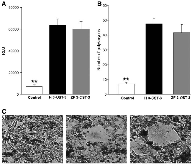
Fig. 3 (A) ZF 3-OST-3-expressing target CHO-K1 cells gain the ability to fuse with effector cells coexpressing HSV-1 glycoproteins gB, gD, gH, and gL. The target cells were transfected with plasmids expressing H 3-OST-3 and ZF 3-OST-3 (as indicated), and luciferase reporter gene. The effector cells were transfected with HSV-1 glycoproteins gB, gD, gH, and gL, and T7 RNA polymerase. A luciferase reporter assay was performed 24 h after the two cell populations were mixed together. Cell fusion was measured in relative luciferase units (RLUs) using a Sirius luminometer (Berthold Detection System). (A) Expression of ZF 3-OST-3 isoform results in the fusion of CHO-K1 cells with HSV-1-glycoprotein-expressing cells as measured by a luciferase assay system with reporter lysis buffer (Promega). The data shown are the means of triplicate measures and are representative of three independent experiments. Double asterisks indicate significant difference from other treatments ( p < 0.01, t-test); error bars represent SD. (B) Quantitative determination of the number of fused polykaryocytes formed after coculture of target cells expressing either ZF-encoded 3-OST-3 or human-encoded 3-OST-3 receptors with the effector CHO-K1 cells expressing HSV-1 glycoproteins. Target CHO-K1 cells expressing empty vector (pDream2.1) cultured with effector CHO-K1 cells were used as negative controls. The data shown are the means of triplicate measures with three independent experiments. Double asterisks indicate significant difference from other treatments ( p < 0.01, t-test); error bars represent SD. (C) Multinucleated cells or polykaryocytes were microscopically observed with cells expressing H 3-OST-3 (panel b) and/or ZF 3-OST-3 (panel c), but not with cells expressing control vector pDream2.1 (panel a). CHO-K1 target cells transfected with plasmids expressing H 3-OST-3 or ZF 3-OST-3 were mixed with the HSV-1 glycoprotein expressing effector cells, and stained with Giemsa at 24 h postmixing. The effects of H 3-OST-3- or ZF 3-OST-3-expressing CHO-K1 cells on multinucleated polykaryocytes formation were observed. Shown are photographs of representative cells (Nikon) after 24 h.