
PUBLICATION
Complex cell rearrangements during intersegmental vessel sprouting and vessel fusion in the zebrafish embryo
- Authors
- Blum, Y., Belting, H.G., Ellertsdottir, E., Herwig, L., Lüders, F., and Affolter, M.
- ID
- ZDB-PUB-080326-18
- Date
- 2008
- Source
- Developmental Biology 316(2): 312-322 (Journal)
- Registered Authors
- Affolter, Markus, Belting, Heinz-Georg Paul (Henry), Ellertsdottir, Elin
- Keywords
- Angiogenesis, ISV, Vessel fusion, VE-cadherin, Adherens junctions, Transgenic, ZO-1, Zebrafish
- MeSH Terms
-
- Animals, Genetically Modified
- Zebrafish/embryology*
- Zebrafish/genetics
- Embryo, Nonmammalian/cytology*
- Embryo, Nonmammalian/physiology*
- PubMed
- 18342303 Full text @ Dev. Biol.
Abstract
The formation of intersegmental blood vessels (ISVs) in the zebrafish embryo serves as a paradigm to study angiogenesis in vivo. ISV formation is thought to occur in discrete steps. First, endothelial cells of the dorsal aorta migrate out and align along the dorsoventral axis. The dorsal-most cell, also called tip cell, then joins with its anterior and posterior neighbours, thus establishing a simple vascular network. The vascular lumen is then established via formation of vacuoles, which eventually fuse with those of adjacent endothelial cells to generate a seamless tube with an intracellular lumen. To investigate the cellular architecture and the development of ISVs in detail, we have analysed the arrangement of endothelial cell junctions and have performed single cell live imaging. In contrast to previous reports, we find that endothelial cells are not arranged in a linear head-to-tail configuration but overlap extensively and form a multicellular tube, which contains an extracellular lumen. Our studies demonstrate that a number of cellular behaviours, such as cell divisions, cell rearrangements and dynamic alterations in cell-cell contacts, have to be considered when studying the morphological and molecular processes involved in ISV and endothelial lumen formation in vivo.
Genes / Markers
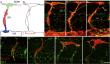
Figure Gallery (6 images)


Expression
Phenotype
Mutations / Transgenics
Human Disease / Model
Sequence Targeting Reagents
Fish
Orthology
Engineered Foreign Genes
Mapping