IMAGE

Fig 3

ID
ZDB-IMAGE-250705-12
Source
Figures for Rissone et al., 2025
Image
Figure Caption

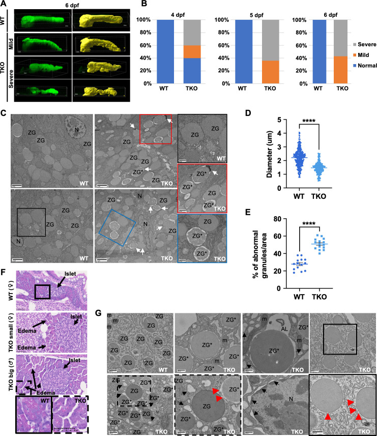
Fig 3 Pancreatic defects observed in TKO embryos and adult fish.

(A) Representative max-projections (green) and Imaris 3D reconstructions (yellow) of confocal Z-stacks of 2CLIP WT and TKO embryos at 6 dpf showing the different classes of phenotypes of the mutant embryos. Dorsal views, anterior to the left. Scale bars, 30 μm. (B) Quantification of the % of pancreas phenotypes observed in WT and TKO embryo populations during development from 4 to 6 dpf. (C) Representative EM pictures comparing acinar cells in WT and TKO 5 dpf embryos. White arrows: zymogen granules in autolysomes. Scale bars, 2 μm. Inset scale bars: 0.8 μm. (D-E) Quantification of zymogen granules size (D) and quality (E) in WT and TKO embryos at 5 dpf. The data represent means ± SEM. Statistical significance was determined by unpaired two-tailed t-test. **** < 0.0001. (F) H&E histological analysis of 4 months old WT and TKO adult fish showing mild signs of pancreatic inflammation (edema, eosin-neg zymogen granules). Insets are showing the edema and the lack of eosin positivity in the TKO mutant pancreas more in detail. Scale bars, 50 μm. (G) Representative EM images of pancreatic tissues from adult WT and TKO fish. Scale bars: 800 and 400 nm. Black arrows mark dilated endoplasmic reticulum and nuclear envelop; red arrowheads label ruptures in the zymogen granule membrane. AL, autolysosome; m, mitochondria; N, nucleus; ZG, zymogen granule; ZG*, abnormal granules.

Figure Data
Acknowledgments
This image is the copyrighted work of the attributed author or publisher, and ZFIN has permission only to display this image to its users. Additional permissions should be obtained from the applicable author or publisher of the image. Full text @ PLoS Genet.