IMAGE

Fig. 1.

ID
ZDB-IMAGE-250701-13
Source
Figures for Lee et al., 2025
Image
Figure Caption

Fig. 1.

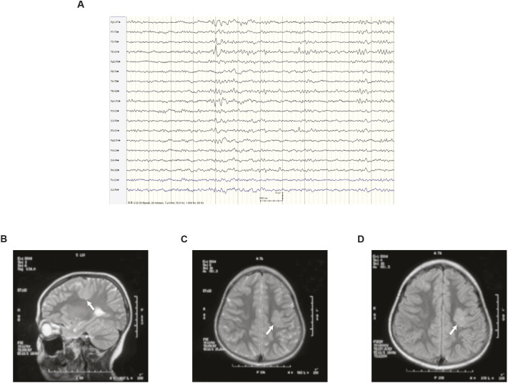
Clinical profiles of an epilepsy patient with SLC25A22 mutation. (A) An electroencephalogram of the patient at the age of 24. (B-D) Brain magnetic resonance imaging of the patient at the age of 9. T2-weighted sagittal (B), axial (C) and fluid-attenuated inversion recovery (FLAIR; D) images are shown. White arrows indicate the left cortical heterotopia.

Acknowledgments
This image is the copyrighted work of the attributed author or publisher, and ZFIN has permission only to display this image to its users. Additional permissions should be obtained from the applicable author or publisher of the image. Full text @ Dis. Model. Mech.