IMAGE

Fig 4

ID
ZDB-IMAGE-250620-9
Source
Figures for Ma et al., 2025
Image
Figure Caption

Fig 4 
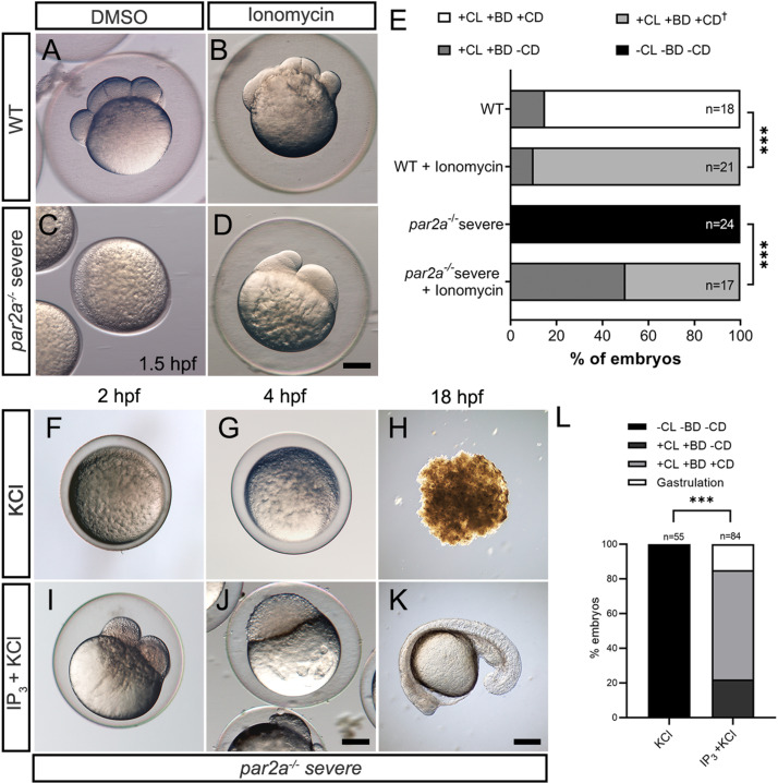
Increasing intracellular Ca2+ either by Ionomycin or IP3 rescues egg activation defects in par2a mutants.

A–D: Images of WT (A, B) or severe par2a mutants (C, D) embryos derived from natural spawning and treated with 0.1% DMSO (A, C) or 2 µM Ionomycin (B, D). E: Proportion of WT and severe par2a−/− embryos showing egg activation and blastomere division phenotypes at 1.5 hpf following treatment with 0.1% DMSO or 2 µM Ionomycin. Key: CL: Chorion Lift, BD: Blastodisc, CD: Cell Division, +: Present, −: absent. : Abnormal. Chi-squared analyses; *** = p < 0.001. F–K: Images of naturally fertilized severe par2a mutant embryos injected with KCl alone (F, G, H) or with IP3 in KCl (I, J, K) at 2 hpf (F, I), 4 hpf (G, J) and 18 hpf (H, K). L: Proportion of egg activation and blastomere division phenotypes in fertilized severe par2a mutant eggs injected with IP3 in KCl or KCl alone. Key: CL: Chorion Lift, BD: Blastodisc, CD: Cell Division, +: Present, −: absent. Chi-squared analyses; *** = p < 0.001. Scale bars: D, J, K = 200 µm. See file S1 Data for underlying data. IP3 is necessary for zebrafish egg activation.

Figure Data
Acknowledgments
This image is the copyrighted work of the attributed author or publisher, and ZFIN has permission only to display this image to its users. Additional permissions should be obtained from the applicable author or publisher of the image. Full text @ PLoS Biol.