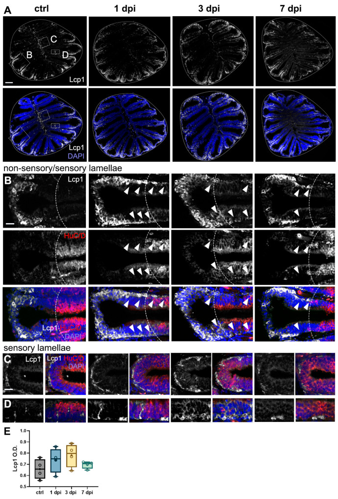
Leukocytic migration within the OE caused by 6-OHDA injections in the OB. (A–D) Lymphocyte cytosolic protein 1 (Lcp1) immunohistochemistry in sections of the OE from controls, 1 dpi, 3 dpi, and 7 dpi. Whole OE (A) and magnified views of the following: (B) distal epithelial fold region, where the OSN-containing sensory region is indicated by HuC/D staining and a dotted line; (C) medial sensory region (HuC/D+) adjacent to the central raphe; and (D) sensory epithelium (HuC/D+). Migration of Lcp1+ cells is indicated by white arrowheads. White: Lcp1; blue: DAPI. Scale bars: 100 µm in (A); 20 µm in (B–D). (E) Quantification of Lcp1+ cells in OE sections from (A) (n = 4). ANOVA: F (3, 12) = 1.42, p = 0.2840. Box plots indicate mean (+), quartiles (boxes), and range (whiskers).
Acknowledgments
This image is the copyrighted work of the attributed author or publisher, and
ZFIN has permission only to display this image to its users.
Additional permissions should be obtained from the applicable author or publisher of the image.
Full text @ Int. J. Mol. Sci.
Your Input Welcome
Thank you for submitting comments. Your input has been emailed to ZFIN curators who may contact you if
additional information is required.
Oops. Something went wrong. Please try again later.