IMAGE

Figure 4

ID
ZDB-IMAGE-250530-20
Source
Figures for Vorhees et al., 2025
Image
Figure Caption

Figure 4

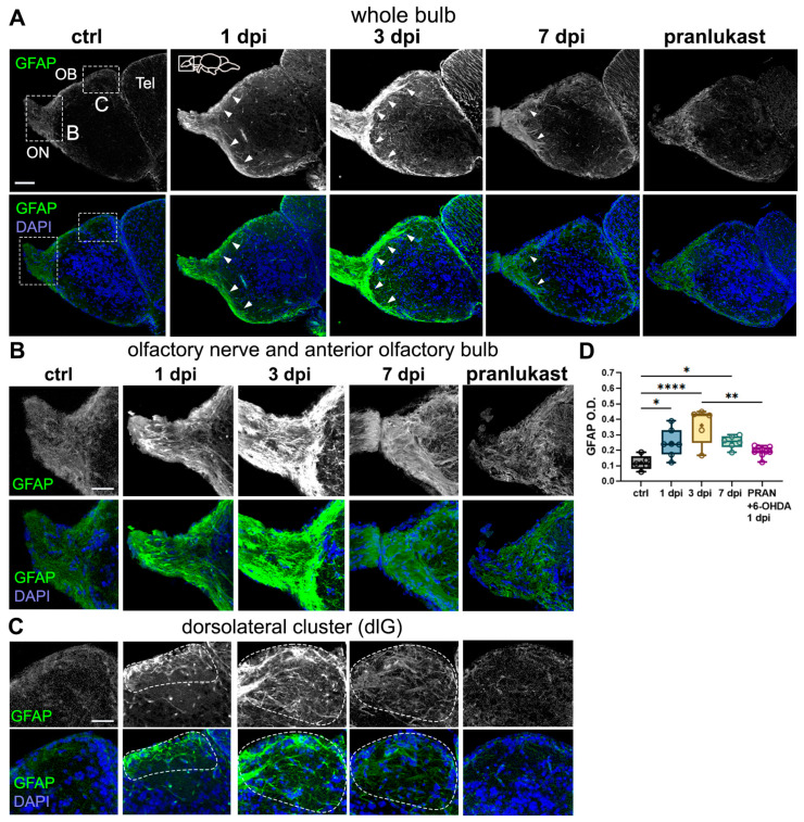
6-OHDA injections lead to astroglial activation in the OB. (AC) Glial fibrillary acidic protein (GFAP) immunohistochemistry in sagittal sections of the OB from controls, 1 dpi, 3 dpi, 7 dpi, and 6-OHDA with the anti-inflammatory drug pranlukast (PRAN). Whole bulb (A) and magnified views of: (B) the olfactory nerve and anterior OB, and (C) the dorsolateral (dlG) glomerular cluster (indicated by dotted lines). GFAP+ staining along the olfactory nerve layer (ONL) is indicated with white arrowheads. Green: GFAP; blue: DAPI. Scale bars: 100 µm in (A); 50 µm in (B,C). (D) Quantification of GFAP O.D. in OB sections from (A) (n = 5–9). ANOVA: F (4, 27) = 8.478, p = 0.0001. ctrl vs. 1 dpi p = 0.0306, ctrl vs. 3 dpi p < 0.0001, ctrl vs. 7 dpi p = 0.0275. 1 dpi vs. 3 dpi p < 0.0001. 3 dpi vs. PRAN p = 0.0017. Box plots indicate mean (+), quartiles (boxes) and range (whiskers). One-way ANOVA, * p < 0.005, ** p < 0.001, **** p < 0.0001.

Acknowledgments
This image is the copyrighted work of the attributed author or publisher, and ZFIN has permission only to display this image to its users. Additional permissions should be obtained from the applicable author or publisher of the image. Full text @ Int. J. Mol. Sci.