
|
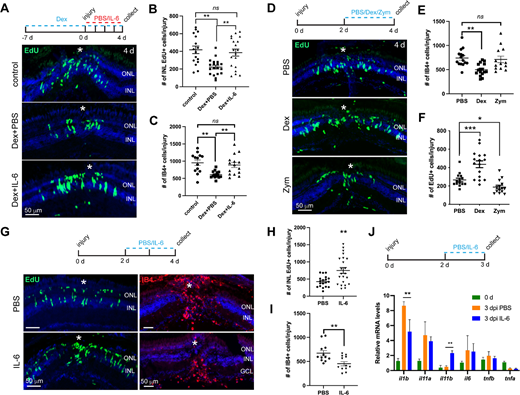
Fig. 5 IL-6 may regulate MGPC formation through phase-dependent pro-inflammatory and anti-inflammatory mechanisms. (A) The experimental timeline, and EdU immunofluorescence showing the formation of MGPCs (INL EdU+ cells) in control or Dex-treated retinas at 4 dpi that received daily PBS- or IL-6 injection. (B) Quantification of the number of INL EdU+ MGPCs per injury in A. (C) Quantification of the number of IB4+ microglia at the injury site in retinas that received the same treatment of A at 2 dpi. (D) The experimental timeline, and EdU immunofluorescence showing the formation of MGPCs at 4 dpi in the retinas that received intravitreous injection of PBS, Dex, or Zym. (E) Quantification of the number of IB4+ microglia at the injury site in the retina in D at 4 dpi. (F) Quantification of the MGPC number per injury at 4 dpi in D. (G) The experimental timeline, and immunofluorescence showing the INL EdU+ MGPCs and IB4+ microglia at 4 dpi in retinas that received intravitreous injection of PBS or zebrafish IL-6 protein (100 ng). (H, I) Quantification of the number of MGPCs and IB4+ cells per injury in G at 4 dpi. (J) The experimental timeline, and qPCR analysis of the mRNA expression of indicated cytokines at 3 dpi in retinas that received intravitreous injection of PBS or zebrafish IL-6 protein (100 ng). White *, site of the stab injury. *, P < 0.05; **, P < 0.01; ***, P <0.001; Dex, dexamethason; GCL, ganglion cell layer; INL, inner nuclear layer; ns, non-significant; ONL, outer nuclear layer; Zym, zymosan A.