
|
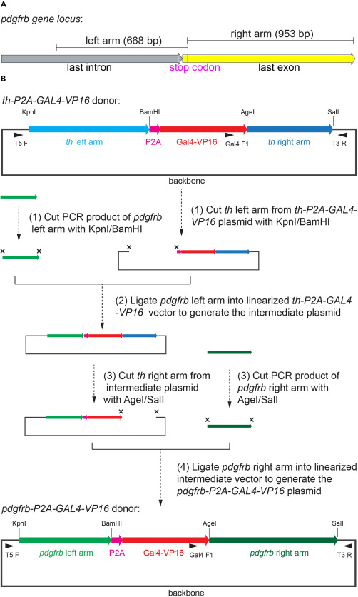
Fig. 3 Schematic representation for the generation of pdgfrb-P2A-Gal4-VP16 donor plasmid (A) Schematic representation for the sequences of the left and right arms for the pdgfrb-P2A-Gal4-VP16 donor plasmid in pdgfrb gene locus. The sequence of last intron, last exon and stop codon are marked. (B) Schematic representation of the flow for generating pdgfrb-P2A-Gal4-VP16 donor plasmid. Use the th-P2A-Gal4-VP16 plasmid as backbone, replace the left arm and right arm orderly. The compositions of pdgfrb-P2A-Gal4-VP16 and pdgfrb-P2A-Gal4-VP16 plasmids are shown. The target sites of the restriction endonucleases KpnI, BamHI, AgeI, and SalI are marked. The binding sites of the primers T5F, Gal4 F1, and T3R are marked.