
|
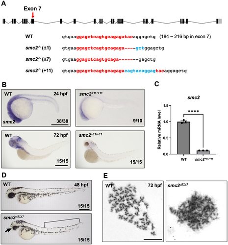
Fig. 2 Generation of smc2 mutants via CRISPR/Cas9 mutagenesis. (A) A diagram of smc2 targeted by a sgRNA at exon 7 (a red arrow) and the sequences of three different smc2 mutant alleles generated using CRISPR/Cas9 mutagenesis. Numbers in parentheses with WT sequences indicate the position in the open reading frame of the targeted exon 7 of smc2. The sgRNA sequence is shown in red bold letters. Deletions are illustrated as dashed lines and insertions as blue letters. (B) Whole-mount in situ hybridization (WISH) using a smc2 probe of smc2 mutant embryos and its WT sibling controls at 24 and 72 h post fertilization (hpf). The numbers shown at the bottom of the images represent the count of representative outcomes observed relative to the total number of embryos obtained. Data from two independent experiments. Scale bar = 400 μm. (C) Quantitative PCR (qPCR) analysis assessing the relative expression levels of the smc2 gene in WT and smc2 null mutant zebrafish embryos at 72 hpf. Expression levels of smc2 were normalized to β-actin, with results presented as relative fold changes. Data were quantified and expressed as the mean ± standard error of the mean (SEM) from three independent biological replicate experiments. Statistical analysis was performed using a t-test to calculate p-values. (****p < 0.0001). (D) Lateral view of smc2 mutant embryos and WT controls at 48 hpf. smc2 mutants demonstrated morphological defects in the head (black arrow) and yolk extension (black square bracket). Data from two independent experiments. Scale bar = 400 μm. (E) Chromosome-spreading assay of WT and smc2 mutant embryos at 72 hpf from three independent experiments performed. The scale bar indicates the designated length in the images. Scale bar = 10 μm.