IMAGE

Fig. 1.

ID
ZDB-IMAGE-250422-101
Source
Figures for Laverde et al., 2025
Image
Figure Caption

Fig. 1.

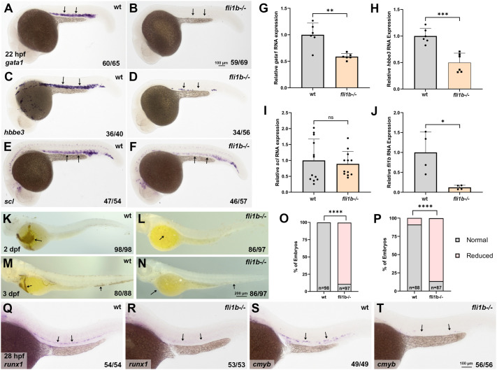
fli1b mutants display reduced primitive and definitive hematopoiesis. (A-F) Whole-mount in situ hybridization (WISH) analysis of expression of the erythroid markers gata1 (A,B) and hbbe3 (C,D) and the hematopoietic marker scl (E,F) in fli1b−/− mutant and wild-type (wt) embryos (in fli1a:GFP background) at 22 h post fertilization (hpf). fli1a:GFP embryos were used as controls to enable fluorescence analysis of any potential defects in vascular development in fli1b−/− embryos, which show GFP expression linked to fli1b mutation. Note greatly reduced expression of gata1 and hbbe3 and moderately reduced expression of scl in the intermediate cell mass region (arrows) in fli1b mutants. (G-J) qPCR analysis of gata1 (G), hbbe3 (H), scl (I) and fli1b (J) expression in fli1b−/− mutant and wt fli1a:GFP embryos at the 20-somite stage. Note the significant reduction in gata1, hbbe3 and fli1b expression in fli1b mutant embryos. Bars show mean±s.d. ns, not significant; *P<0.05; **P<0.01; ***P<0.001; Student's two-tailed unpaired t-test. (K-N) Heme staining using o-dianisidine in fli1b mutant and control wt fli1a:GFP embryos at 2 and 3 days post fertilization (dpf). Note greatly reduced staining (arrows) in fli1b mutant embryos. (O,P) Quantification of embryos with normal and reduced heme staining at 2 dpf (O) and 3 dpf (P). ****P<0.0001; Fisher's exact test. (Q-T) WISH analysis for expression of the hematopoietic stem cell and progenitor markers runx1 and cmyb at 28 hpf in the trunk region of fli1b mutant and control wt fli1a:GFP embryos. Note the greatly reduced or absent staining (arrows) in fli1b mutants. All experiments have been replicated at least twice.

Acknowledgments
This image is the copyrighted work of the attributed author or publisher, and ZFIN has permission only to display this image to its users. Additional permissions should be obtained from the applicable author or publisher of the image. Full text @ Biol. Open