
Image
|
Figure Caption
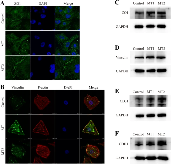
Fig. 6 Cell adhesion proteins are upregulated following CRIP2 depletion. (A–B) ZO1 and Vinculin expression in the control and CRIP2-MT cell lines were determined by immunostaining. TRITC-phalloidin and DAPI were used to stain F-actin and the nucleus, respectively. (C-F) ZO1, vinculin, CD31 and CDH1 expression in the control and CRIP2-MT cell lines, as determined by Western blotting
Acknowledgments
This image is the copyrighted work of the attributed author or publisher, and
ZFIN has permission only to display this image to its users.
Additional permissions should be obtained from the applicable author or publisher of the image.
Full text @ Cell. Mol. Life Sci.